Screen share
Upload case
Log in
Search
in
ALL EXAMS
Show images
FASTEST SPOT Case 10 Axial CTA
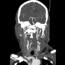
FASTEST SPOT Case 19 Cor CTA (EXAMPLE)
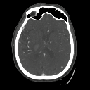
FASTEST SPOT Case 19 Axial CTA (EXAMPLE)
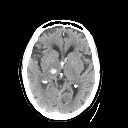
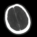
FASTEST SPOT Case 19 NCCT (EXAMPLE)
FASTEST SPOT Case 18 Cor CTA
FASTEST SPOT Case 18 Axial CTA
FASTEST SPOT Case 18 NCCT
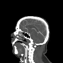
FASTEST SPOT Case 15 Sag CTA
FASTEST SPOT Case 15 Axial CTA
FASTEST SPOT Case 15 NCCT
FASTEST SPOT Case 14 Cor CTA
FASTEST SPOT Case 14 Axial CTA
FASTEST SPOT Case 14 NCCT
FASTEST SPOT Case 13 Cor CTA
FASTEST SPOT Case 13 Axial CTA
FASTEST SPOT Case 13 NCCT
FASTEST SPOT Case 12 Cor CTA
FASTEST SPOT Case 12 Axial CTA
FASTEST SPOT Case 12 NCCT
FASTEST SPOT Case 10 Sag CTA
FASTEST SPOT Case 10 Cor CTA
FASTEST SPOT Case 10 NCCT
FASTEST SPOT Case 9 Cor CTA
FASTEST SPOT Case 9 Axial CTA
FASTEST SPOT Case 9 NCCT
FASTEST SPOT Case 8 Cor CTA
FASTEST SPOT Case 8 Axial CTA
FASTEST SPOT Case 8 NCCT
Spasm CT
Calcified M3
M3 Perf
A3 and fu
A3 Occlusion Far
CT P2 FU
P2 Left Occlusion MRI
P2 Left Occlusion
P2 Example with FU CT
P2 MRI
P2 Occlusion Causing Homonymous Hemianopia
Large AT Anterior Temporal (M1 occlusion)
Functional M1 2M2
A3 Occlusion
MRI A3
M3 Brocas MRI
M3 Anterior Branch to Broca's - Too small and high risk of rupture
Dominant M2 - Do not enroll
Perforation Complication During M2 anterior branch
M3 MRI
M3 to Motor Strip MeVO
A2/3 Occlusion
M3 Occlusion CT - 01-051.
M3 Occlusion MRI - 01-051
M3 Occlusion CT - 01-051
double mevo MRI
double double
Mobile Clot
Engulfing Clot BadASS
Blind Exchange Mini Pinning
Bovine 5 - AG
Bovine 6 - SC
Bovine 7 - LM
Bovine Arches 2 - AB (AP Lateral LAO) 1
Bovine Arch 8 (AP)
Bovine Arches 3 - ZA
Bovine Arches 1 - (EF AP Lateral LAO)
Bovine Arches 4 - BO ((AP, Lateral, LAO)
Tough Arch 2 (AP, Lateral, LAO)
Tough Arch 1 (AP, Lateral, LAO)
FASTEST 10
FASTEST 9
FASTEST 8
FASTEST 7
FASTEST 6
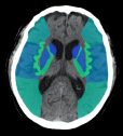
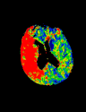
Aspects_Regions Color Petra
FASTEST Example
FASTEST 5
FASTEST 4
FASTEST 3
FASTEST 2
FASTEST 1
colorviz alone
ColorViz
Post MRI
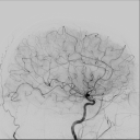
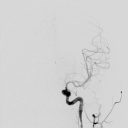
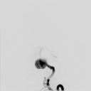
Angio
Bilateral carotid dissection MRI
Bilateral Carotid Dissections
Non-ketotic hyperglycemic hemichorea (NHH)
mCTA - ACA Occlusion
Pontine Recurrent Infarcts
mCTA PCA Occlusion
LV Aneurysm with thrombus
077 6 0 0 0 1 0 0 0 1 1 1
037 5 0 1 0 0 0 1 0 1 1 1
042 5 1 1 0 1 0 0 0 1 1 0
162 5 0 0 0 1 0 1 0 1 1 1
210 5 0 0 0 1 0 1 1 1 1 0
314 9 0 0 0 0 0 0 0 1 0 0
065 8 1 1 0 0 0 0 0 0 0 0
408 8 1 1 0 0 0 0 0 0 0 0
267 8 1 1 0 0 0 0 0 0 0 0
038 8 0 1 0 1 0 0 0 0 0 0
074 7 1 1 0 0 0 0 0 0 1 0
040 2 1 1 0 1 0 1 1 1 1 1
024 3 0 0 0 1 1 1 1 1 1 1 fetal origin of the left PCA since subacute infarct of the left MCA AND PCA territory
369 3 0 1 0 1 0 1 1 1 1 1
064 3 1 1 0 1 0 1 0 1 1 1
342 4 0 1 0 1 1 1 1 0 0 1
027 5 1 1 0 1 0 1 1 0 0 0
162 5 0 0 0 1 0 1 0 1 1 1
210 5 0 0 0 1 0 1 1 1 1 0
148 6 1 1 0 1 0 1 0 0 0 0
099 6 0 1 0 1 0 0 0 1 1 0
078 6 0 1 0 1 0 0 0 1 1 0
042 6 0 0 0 1 0 0 0 1 1 1
Right insular infarct - findings
Terson's syndrome
M2 MRI
M3 mCTA Occlusion
M3 Intervention in high NIHSS
Fat Embolism MRI
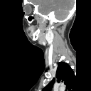
CT Fat embolism syndrome
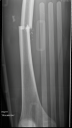
xray femur fracture
JNA MRI
JNA CT
Carotid body paraganglioma.
ECA and Ascending Pharyngeal Artery
Vidian Artery from ICA Petrous Segment
Diffuse hypoxic ischemic injury post amphetamines HIE adult diffuse
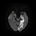
Tiny_Brainstem_Lateral_Medulla_Infarct_DWI_subtlety
Curved MPR and Needle View
Corticolaminar Necrosis
Extensive Dural AVF
Mitral Valve Calcification as source of embolus
Aortic Dissection as Cause of Stroke
Watershed MRI
CapsuloStriatal Infarct
Loeys-Dietz syndrome (LDS) Vessels
Luxury perfusion - bad no scale - post resolution of cloth in patient with severe aphasia
Innonimate Stenosis and mcta delay, subclavian steal and collaterals
Left PCA occlusion
head of caudate infarct and ipsilateral ventricular enlargement
Anatomy 3rd ventricle
Stasis in Carotid Web
Accessory MCA
chiari I
Double SCA AICA and Percheron
Chasing the Dragon (diff for leukoencephalopathy)
Angio Anatomy - ECA low
Angio Anatomy - ECA high
Angio Anatomy - RCA VERT
Angio Anatomy - Lateral only
MRI for CTP correlation
Spot Sign CTA
Anatomy 3rd ventricular anatomy Choroid Plexus
Multiple Carotid Dissection
CTP and miStar Utility question and wrong on infarct
Tortous kissing carotids, multiphase delay
mCTA for Spot Sign Vessel vs hematoma
avm vert left vert run - surprise not aneurysm
vert avm cta
avm fed from ECA
Right M3 Cloth and Subtle Paynchemal GW loss
dAVF - CT and CTV
dAVF Angio
MR Angio Anatomy: 3T normal peds
Excellent collaterals on mcTA
cerebellar strok e and hydro
Early Cortical G/W Differentiation Loss and Left M2 cloth
icAD intracranial athero
Carotid T Occlusion CTP
motor strip stroke XA angio
motor strip stroke mri
motor strip stroke mcta - good delay
ACA CT F/U first phase finding not seen on second delay
ACA MRI first phase finding not seen on second delay
Carotid T Occlusion mcta
Tuberous Sclerosis TS right
Tuberous Sclerosis TS
ACA mCTA first phase finding not seen on second delay
Tuberous Sclerosis (TS)
Paraophthalmic Aneurysm Angio
Angio Paraophthalmic 3D
Subdraul Extension of SAH post Anu Rupture
Paraophthalmic aneurysm - Angio 3D
Crossed Cerebellar Diaschesis SPECT
Superior Cereberral Infarct CT - Crossed Cerebellar Diaschesis
Superior Cereberral Infarct - Crossed Cerebellar Diaschesis
090 9 M3 2 2 2 2 2 2 2 2 2 2 20 5 0
077 9 M1 2 2 2 1 2 2 2 2 2 2 19 4 1
060 10 0 2 2 2 2 2 2 2 2 2 2 20 5 0
050 8 M1 2 2 1 0 2 1 2 2 2 2 16 3 1
049 7.5 M2 2 1 0 0 2 2 2 2 2 2 15 4 1
041 10 0 2 2 2 2 2 2 2 2 2 2 20 5 0
040 9.5 0 2 2 2 2 2 2 2 2 2 2 20 5 0
030 10 ICA 2 2 2 1 2 2 2 2 2 2 19 5 1
Cortical Vein CT
Cortical Vein MRI
Cortical Vein Thrombosis
029 1 1 1 M2 4 9.5 19 2 1 2 2 2 2 2 2 2 2 1
027 2 2 1 M1 4 7.5 15 2 1 0 0 2 2 2 2 2 2 1
024 2 2 1 M1 4 4 8 2 1 1 0 1 0 0 0 1 2 0
020 1 1 0 M3 5 9 18 2 2 2 1 2 2 2 2 1 2 0
019 2 3 1 M2 5 9 18 2 2 1 1 2 2 2 2 2 2 1
Numero1
mCTA Subtle Hangup in Pt with left facial droop and arm and leg weakness
DWI for Subtle mCTA pickup
PCA and MCA delay on mCTA
Papez Circuit
Left PCA and MCA delay on mCTA
Hippocampal Infarct
Basilar Thrombus
Basilar Thrombus Hyperdense NCCT
Tight Left MCA Occlusion and watershed infarct. rapid onset
Cervical ICA Dissection
Cervical ICA Dissection
CTP - No pneumbra
Black Hole collaterals
Stroke Mimic - Left Parietal Mass
SAH at the gym in young female
MRI Pre HOD
MRI HOD
Tectal AVM
Artery of Percheron Angio
Anoxic CTP
Anoxic CT
MS with Optic Neuritis in a gentleman
Bilat Occipital infract mcta proper
SCA Aneurysm
Thoracic Spinal AVM
PICA Occlusion
Bilat PCA MRI
Bilateral Occipital Infarcts - CTP
PCA Territory on mCTA
Subependymal Nodular Heterotopia Causing Seizures
Non Occlusive Subtle Thrombus Angio XA
Non Occlusive Subtle Thrombus CTP
Moya Moya Angio
Moya Moya CT and CTA
Persistant Trigeminal Artery and Fetal PCA - Saltzman classification
Pancoast Tumor Scout
PICA Left infarct
Butterfly GBM
Atrial Myxoma Cause for Stroke
Thick MIP Useful
Giant ACA Aneurysm
PICA Aneurysm
CT early signs basics
Traumatic CC CT Initial
Traumatic CC MRI - right brainstem infarct
Immediate Improvement XA
Immediate Improvement MR
Immediate Improvement CTA
XA - Attempt to restore
Followup Poor Outcome
NPH with DESH
Mid Basilar Aneurym XA
R MCA Clacified Emblus - What to do?
HII - Hypoxic Ischemic Injury - Diffuse and Very Subtle
Herpes Encephalitis - Left temporal lobe subtle
Hemorrhagic Transformation XA
Hemorrhagic Transformation f/u CT
Hemorrhagic Transformation after Free thrombus CT
Hemorrhagic Transformation MRI - nice case
vr test
Tortous Carotid XA
Tortous Carotid MRI
Amyloid with perimedullary venous calcificaitons CT
Amyloid with perimedullary venous calcificaitons
no ncct change,XA poor collateral Anterior choroidal. Terminus. Captive cap6 87 walker before.
no ncct change,MRI poor collateral Anterior choroidal. Terminus. Captive cap6 87 walker before.
no ncct change, poor collateral Anterior choroidal. Terminus. Captive cap6 87 walker before.
aspects 8 great XA
aspects 8 mri
aspects 8
T anterior coroidal new occlusion XA
T anterior coroidal new occlusion MRI
T anterior coroidal new change and recanalized pcomm fetal
T anterior coroidal
Left M1 with Good Collaterals FU CT
Left M1 with Good Collaterals
Carotid stent
Post EVT Injury Carotid Stent / Hemmohragic transformation
left vert cta
left vert mri
Right ACA M3 mCTA Occlusion -fu ACA Infarct
53 year old female with acute left hemiparesis XA
53 year old female with acute left hemiparesis, mri
53 year old female with acute left hemiparesis, hemineglect and dysarthria. Right MCA
Right ACA M3 mCTA Occlusion - 77 year old female presented with acute left leg weakness.
44 year old male with acute onset speech difficulty ctp
44 year old male with acute onset speech difficulty mri
44 year old male with acute onset speech difficulty CT
86 year old cta multi
86 year old xa
86 year old mri
86 year old ctp
86 year old
S CT
S MRI
Carotid T Occlusion MRI
Carotid T Occlusion CTA
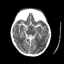
Right CT Scroll Together (keep)
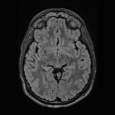
Right MCA MRI
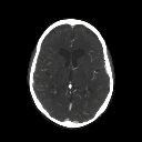
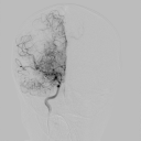
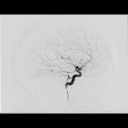
Right MCA CTA (Whole Case)
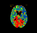
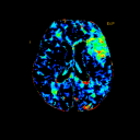
Right MCA Perfusion
Tumor mimic temporal lobe CT
Stroke Mimic Parietal Tumor
Stroke Mimic Parietal Tumor
T bone Coronal for Anatomy
Suppl Coronal T2 for Papez
T Bone for Anatomy
Multi System Atrophy (MSA)
Papez Circuit Anatomy
PRES and Subcortical Ufibers CT
PRES and Subcortical Ufibers
Moya Moya MRI
Moya Moya CT
Moya Moya MRA Single case
davf mr
fetal pcom and posterior R PCA
fetal pcom and posterior R PCA
Calcified emboli from carotid bifurcation plaque
When to Intervene or Not
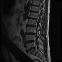
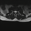
Disk and synovial cyst Spine MRI causing cauda
Inflammatory Polyneuropathy - acute CIDP
Calcified Embolus CT
Calcified Embolus MRI
MRI - Right M1 hyperdense with moderate collaterals
Right M1 hyperdense with moderate collaterals Scroll
Right M1 hyperdense with moderate collaterals Series (nonscroll)
Distal occlusion, use of mCTA - MRI
Case 5 - Right MCA - Collateral
Case 4 Very large Right MCA
Case 3 Left M3 Cloth
Case 2 mCTA
Case 1 ASPECTS R-MCA
ASPECTS Calculations
Single Axial Plain CT Large R MCA Stroke - mustache sign
AVM Case - nicely annotated and full story
Showing all 339 matches
ALL EXAMS
FOLDERS
Arch Anatomy
ASPECTS Scoring
FASTEST
Neuro Cases
Stroke Cases
Vascular Anatomy